
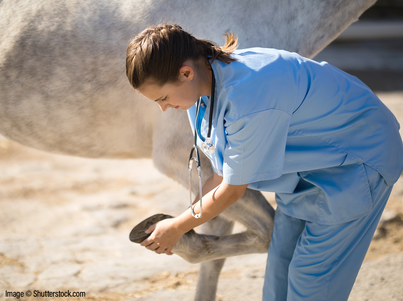
Horses coming in for lameness investigations at the clinic may have several different procedures undertaken to try and pin-point the cause of lameness. These commonly include:
- Physical examination
- Nerve blocks
- Joint blocks
- Radiography
- Ultrasound.
Trotting Up
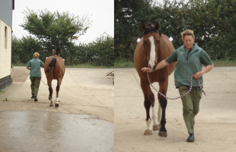
Initially horses are examined before any nerve blocks are placed.
We commonly trot horses up, perform flexion tests, lunge in the school and on a hard surface. More subtle lamenesses may require the horse to be worked under saddle for a time. We can then grade the horse’s lameness for each leg affected.
This is used as a comparison when later nerve blocks are used.
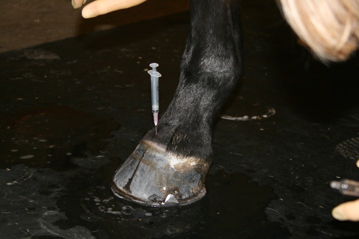
Nerve Blocks
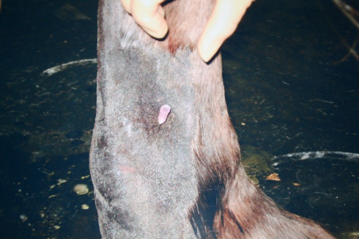
Since the horse cannot tell us exactly which part of the leg is hurting, we have to use another method to deduce which area we need to take a closer look at.
Local anaesthetic is placed around the nerves running up the horse’s leg, at strategic points to ‘block’ the nerve signal (numbs the area).
When the nerves coming from the painful area are ‘blocked’ the horse should look ‘magically cured’ – or at least much less lame when re-assessed!
We start at the foot end and work up the leg. This way, the leg is divided into smaller ‘zones’ of interest.
Radiography and Ultrasonography
Once we have narrowed the lameness down to an area of interest, we normally find x-rays and/or ultrasound are helpful to diagnose the problem.
Joint Bocks
Local anaesthetic can be injected under sterile conditions into the joint to desensitise it. The horse is then assessed again to judge the effect.
Joint blocks normally have to be performed on a separate occasion to nerve blocks, to give the horse time for sensation to have come back to the leg fully so as not to give confusing results.
Joint blocks carry a very small risk of introducing infection into the joint – which is a serious problem. Therefore we clip a small area over the joint and prepare as if for surgery before injecting sterile local anaesthetic.
We sometimes prepare for nerve blocks in the same way if we are injecting very close to a joint.
Referrals
Occasionally a horse will need to be referred to a specialist centre to complete the lameness work-up. Specialist procedures can include ‘bone-scanning’ (scintigraphy) or MRI.